
Using powerful microscopy techniques, a research team led by scientists at the National Institute on Deafness and Other Communication Disorders (NIDCD), part of the National Institutes of Health (NIH), has pinpointed in mice the precise cellular location of two proteins known to be important for hearing and balance. The discovery provides additional evidence that the proteins, TMC1 and TMC2, are part of the channel complex that is essential for the inner ear to process sound and the signals that are key to balance.
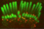
Immunofluorescence of TMC2 (green) in confocal images of mice inner hair cell stereocilia (red).
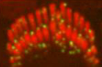
Immunofluorescence of TMC1 (red) in confocal images of rat inner hair cell and outer hair cell stereocilia (green).
The study appeared in the September 8 edition of Cell Reports, and was co-led by Bechara Kachar, M.D., chief of the NIDCD's Laboratory of Cell Structure and Dynamics, and NIDCD Scientific Director Andrew J. Griffith, M.D., Ph.D. The findings expand knowledge of the structure of the channel, enable scientists to further explore the mechanisms by which it functions, and may lead to new insights into certain hearing and balance disorders.
Hair cells, which are topped by thread-like structures called stereocilia, are the key sensory cells in the inner ear. Mechanoelectrical transduction (MET) channels, which are thought to reside at stereocilia tips, are critical to the cells' function. The channels detect mechanical signals from sound vibrations or changes in our position and convert these signals into electrical pulses that are sent to and interpreted by the brain.
While scientists have learned a lot about how the MET channel functions, its molecular structure largely remains a mystery. TMC1 and TMC2 have long been suspected to be parts of the channel complex for several reasons. The structures of both proteins suggest that they lay in the hair cell's membrane, as would be expected for parts of the molecular channel, and in the absence of the proteins, the MET channel doesn't function normally. In addition, the TMC1 and TMC2 genes are turned on at the same time in development that the MET channel becomes functional. But proving that TMC1 and TMC2 are part of the channel complex—rather than having an indirect effect on its function—has been challenging.
To further investigate the location of TMC 1 and TMC2 in the MET channel, the researchers tagged mouse hair cells with with fluorescent proteins and observed the proteins using high-resolution confocal microscopy. This microscopy technique enables scientists to visualize cells in detail in three dimensions.
The scientists found that TMC1 and TMC2 are found along the length of developing stereocilia. As the hair cells matured, the two proteins localized predominantly to the stereocilia tips. The images also showed that TMC1 and TMC2 are absent from the tips of the tallest stereocilia, where there are no detectable MET channels.
By uncovering the TMC1 and TMC2 localization pattern in hair cells, this work contributes to growing evidence that these proteins are components of the MET channel complex, and that they may be the pore-forming transmembrane proteins of the channel.
This work was supported by NIH/NIDCD intramural research funds (Z01–DC000002 and Z01–DC000060) and by NIH/NIDCD grant R01–DC013521.
- TMC1 and TMC2 Localize at the Site of Mechanotransduction in Mammalian Inner Ear Hair Cell Stereocilia. Kurima K, Ebrahim S, Pan B, Sedlacek M, Sengupta P, Millis BA, Cui R, Nakanishi H, Fujikawa T, Kawashima Y, Choi BY, Monahan K, Holt JR, Griffith AJ, Kachar B. Cell Rep. 2015 Aug 26. pii: S2211–1247(15)00848-7. doi: 10.1016/j.celrep.2015.07.058. [Epub ahead of print] PMID: 26321635